


 Yeast Display VHH Discovery Service
 Immunogen Design & Production
 Premade Library Screening
 Phage Display Nanobody Discovery Service
 Animal Immunization
 Yeast Display Peptide Library Screening

 Nanobody Discovery for ADC
 Nanobody Discovery for Membrane Targets
 Nanobody Discovery for Cell Therapy
 TCR-Mimic Nanobody Discovery


 Antibody Affinity Maturation Service
 Nanobody Humanization Service
How library size affects phage display nanobody discovery is not answered by counting bacterial colonies alone. The practical question is whether a library contains enough unique VHH sequences to represent the target’s available epitopes, while retaining display quality and a workable screening workload. In a phage display nanobody library screening project, researchers must balance library complexity, antigen diversity, panning stringency, CDR3 representation, and downstream validation. An experienced Phage Display VHH Discovery Service, including the workflow offered by 阿帕克, should therefore report both total transformants and measured sequence diversity rather than presenting one large number as proof of performance.
Key answer: a larger library can increase the probability of finding rare binders, but only when the additional members are genuinely different, correctly folded, displayed, and compatible with the screening format. A nominal library of 1010 transformants may contain far fewer unique clones if amplification bottlenecks, recombination, or growth bias have reduced diversity. Conversely, a smaller, high-quality immune or synthetic VHH library can produce useful hits when the antigen, epitope, and panning conditions are well matched.
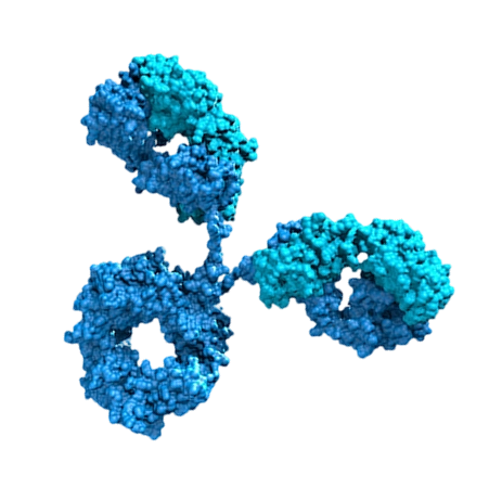
Phage display links a displayed nanobody phenotype to the DNA that encodes it. During biopanning, phage particles are exposed to an immobilized or solution-phase antigen, non-binders are removed, and retained particles are amplified for subsequent rounds. Library size affects the starting probability that a clone with the required binding properties is present.
If a desired clone has a true frequency p in a library and N independent clones are screened, the probability of sampling at least one copy is:
This equation describes sampling probability; it does not prove that every transformed bacterium is unique. For example, if the desired sequence occurs at 1 in 108 independent members, screening approximately 108 independent members gives a substantial chance of sampling it, while screening substantially fewer independent members reduces that chance. The result changes if the clone is toxic, grows slowly, displays poorly, or is lost during amplification.
Why Library Size Matters in Phage Display VHH Discovery Service Projects
- Nominal library size: the number of antibiotic-resistant transformants estimated from dilution plating.
- Independent transformants: the estimated number of separate transformation events represented in the recovered library.
- Unique sequence diversity: the number of distinct VHH nucleotide or amino-acid sequences.
- Functional diversity: the number of clones that fold, display, remain soluble when expressed, and bind under the selected assay conditions.
These values can differ by orders of magnitude. Duplicate sequencing, uneven colony representation, truncations, frame shifts, stop codons, and biased propagation all reduce the effective search space. A library-quality assessment should therefore include colony counting, insert-size analysis, full-length rate, sequence sampling, and a method for estimating redundancy.
Phage Display Nanobody Library Screening: Nominal Size Versus Functional Diversity
The following are published research cases rather than invented customer testimonials. They illustrate why library size must be interpreted together with immunization strategy, VHH architecture, antigen format, and screening design.
Published User Cases That Show the Role of Library Design
Pardon and colleagues published a general protocol for generating nanobodies from immunized camelids and applying phage display to identify antigen binders. Their workflow demonstrates a central point: an immune library does not rely only on random coverage. Antigen exposure produces in vivo enrichment before cloning, so a smaller library with strong antigen-specific representation can be more productive than a larger naïve library for the same target. The study is widely used as a practical reference for llama or alpaca immunization, lymphocyte collection, VHH amplification, phage display, and iterative selection.
Practical lesson: if the project uses an immune library, ask for the immunization antigen, sampling time, lymphoid tissue source, library size, insert quality, and sequence diversity. A large immune library is useful only if the immune response has been captured without excessive PCR or cloning bias.
Case 1: Immunized Camelid Libraries and Antigen-Driven VHH Enrichment
Synthetic antibody and nanobody platforms deliberately control framework sequences and diversify selected complementarity-determining regions, especially CDR3. This approach makes library composition measurable and allows designers to avoid liabilities such as cysteine patterns, unstable frameworks, or excessive hydrophobicity. The search space may be smaller than the theoretical combination of all amino-acid sequences, but its members can be more compatible with expression and folding.
Practical lesson: when comparing two Phage Display VHH Discovery Service providers, request the distribution of CDR3 lengths, amino-acid composition, framework diversity, and the fraction of full-length clones. A claimed library size without this information cannot establish useful diversity.
Case 2: Synthetic and Semi-Synthetic Nanobody Libraries
Published structural biology workflows have used immunized nanobody libraries to isolate binders that stabilize membrane proteins or soluble protein conformations for crystallography and cryo-electron microscopy. In these projects, the desired property is often not merely high affinity. The binder may need to recognize a particular conformational state, avoid competition with a ligand, and form a stable complex suitable for purification. Multiple panning formats and functional screens are therefore used after initial enrichment.
Practical lesson: a very large library does not compensate for an antigen that is misfolded, denatured, incorrectly glycosylated, or presented in the wrong oligomeric state. Antigen quality can be the limiting variable before library size becomes limiting.
Representative sources include Pardon et al., Nature Protocols 2014, “A general protocol for the generation of Nanobodies for structural biology,” and Muyldermans, Annual Review of Biochemistry 2013, “Nanobodies: natural single-domain antibodies.” These publications describe established nanobody-generation principles and should be consulted alongside target-specific literature.
Case 3: Structure-Guided Nanobody Selection
Required Preparation for a Phage Display Nanobody Library Project
- Purified target antigen with documented concentration, purity, buffer, oligomeric state, and activity.
- Positive and negative control proteins, ideally including a close homolog or unrelated protein with similar physicochemical properties.
- Immune, naïve, synthetic, or semi-synthetic VHH phage library with a documented construction method.
- Phage rescue system, host Escherichia coli strain, helper phage or phagemid system, and selective antibiotics.
- Binding plates, magnetic beads, or a solution-phase capture system compatible with the antigen.
- Wash buffer, blocking reagent, elution reagent, PCR reagents, plasmid-miniprep materials, and sequencing access.
- Downstream expression vectors, host cells, protein purification supplies, and an orthogonal binding assay such as ELISA, BLI, SPR, flow cytometry, or microscopy.
Materials and Reagents for Phage Display VHH Discovery Service
- Library size: ask whether the number is based on independent transformants or a theoretical calculation.
- Insert quality: request the percentage of clones containing a full-length VHH insert in the correct reading frame.
- Sequence diversity: request the number of sequenced clones, duplicate rate, framework distribution, CDR3 length distribution, and stop-codon frequency.
- Display format: confirm whether the VHH is displayed as a pIII or pVIII fusion and whether display valency may influence apparent avidity.
- Antigen presentation: define whether selection will use purified protein, cells, virus-like particles, liposomes, or a conformational complex.
- Deliverables: specify whether the final package includes sequences, expression constructs, purified VHH, kinetic data, cross-reactivity testing, and raw sequencing files.
Information to Request Before Choosing a Library
Step-by-Step Workflow for Phage Display Nanobody Library Screening
1. Define the Required Binder Before Measuring Library Size
Tools: target specification sheet, assay protocol, antigen activity assay, and risk assessment.
Action: define whether the required VHH must bind a linear epitope, a conformational epitope, a cell-surface target, a specific species ortholog, or a ligand-bound state. Set acceptance criteria for affinity, kinetics, specificity, expression yield, aggregation, and functional activity.
Parameters: record target concentration range, assay temperature, buffer composition, allowed regeneration conditions, and whether monovalent measurements are required.
Check: confirm that the proposed antigen format preserves the biological state relevant to the final application.
Failure fix: if the antigen loses activity after immobilization, switch to solution-phase panning, capture through a tag positioned away from the binding site, or use cell-based selection.
2. Qualify Library Size and Effective Diversity
Tools: dilution plates, colony counter, insert PCR, agarose gel, Sanger sequencing, and next-generation sequencing when appropriate.
Action: calculate independent transformants from serial dilution plates, test insert presence and size, and sequence a representative clone set before selection.
Parameters: use enough colonies to estimate redundancy rather than sequencing only a few visually different colonies. Report the sampling depth and the method used to cluster identical or closely related sequences.
Check: compare nominal size with full-length insert rate, unique-sequence fraction, CDR3 distribution, and framework balance.
Failure fix: if the library is dominated by duplicates or truncated inserts, rebuild or re-amplify from an earlier low-cycle template instead of relying on repeated panning.
3. Prepare and Test the Antigen
Tools: SDS-PAGE, SEC, mass spectrometry when needed, activity assay, and analytical binding assay.
Action: confirm purity, monodispersity, concentration, and retained activity. For membrane proteins, verify detergent, nanodisc, liposome, or cell-surface presentation.
Parameters: keep the antigen concentration and immobilization chemistry consistent across rounds unless the selection plan intentionally changes them.
Check: test nonspecific binding to the matrix, tag, carrier protein, and blocking reagent.
Failure fix: if background binding is high, add negative selections against the matrix or tag, change the blocking system, reduce exposure time, or use a different capture orientation.
4. Rescue and Normalize the Phage Input
Tools: host cells, helper phage or phagemid system, antibiotic selection, centrifuge, and phage titration assay.
Action: rescue the library under conditions that preserve representation and avoid excessive outgrowth of fast-growing clones.
Parameters: use the supplier’s validated host strain, infection conditions, antibiotic concentrations, and helper-phage protocol. Avoid extending culture time beyond the validated growth window.
Check: determine phage titer and compare input amounts across panning rounds. Monitor whether the library remains infective and whether a control phage behaves as expected.
Failure fix: if infectivity falls, check host competence, helper-phage quality, antibiotic exposure, culture density, and storage history. Do not interpret a low-output round as evidence that the antigen has no binders.
5. Perform the First Panning Round With Diversity in Mind
Tools: antigen-coated surface, magnetic beads, wash buffer, rotator, and elution system.
Action: expose the library to the antigen while running a matched negative-selection control. Separate bound from unbound phage, elute, infect host cells, and retain an aliquot for titer measurement.
Parameters: use a permissive first round to avoid eliminating weak but genuine binders. Record antigen amount, contact time, wash number, wash volume, detergent concentration, and elution conditions.
Check: calculate recovery and compare antigen-positive selection with the negative control.
Failure fix: if recovery is near background, confirm antigen activity, phage infectivity, capture orientation, and elution efficiency before increasing library size.
6. Increase Stringency Gradually in Later Rounds
Tools: the same panning platform, dilution series, competitor protein, and round-by-round sequencing or qPCR monitoring.
Action: apply more stringent washing, lower antigen exposure, soluble competition, off-rate selection, or negative selection against homologous proteins in later rounds.
Parameters: change one major stringency variable at a time so that enrichment can be interpreted. Use a pre-defined stopping rule based on signal-to-background, sequence convergence, or functional activity.
Check: measure enrichment relative to the input and negative control rather than relying on an increasing raw phage count.
Failure fix: if all clones converge to one sticky sequence, reduce amplification bias, add counter-selection, shorten contact time, or restart from an earlier round.
7. Identify Enriched VHH Sequences
Tools: colony PCR, Sanger sequencing, amplicon sequencing, sequence-clustering software, and a VHH annotation tool.
Action: sequence clones from multiple rounds and compare CDR3 sequences, framework usage, stop codons, insertions, deletions, and repeated motifs.
Parameters: sample independent colonies rather than repeatedly sequencing the same clonal streak. Include early and late rounds to distinguish genuine enrichment from amplification artifacts.
Check: confirm that enriched sequences are full-length, in-frame, and not identical because of cross-contamination.
Failure fix: if sequence diversity collapses before functional enrichment, reduce panning pressure, use a different host or rescue format, and inspect growth rates of representative clones.
8. Express Soluble VHH and Confirm Binding Orthogonally
Tools: expression vector, bacterial or mammalian host, affinity purification, SEC, ELISA, BLI or SPR, and an unrelated-protein panel.
Action: express soluble VHH without the phage fusion and test binding using an assay that differs from the discovery format.
Parameters: measure concentration, purity, monomer percentage, apparent affinity, association rate, dissociation rate, and reproducibility across protein preparations.
Check: compare target binding with tag-only, matrix-only, homologous-protein, and unrelated-protein controls.
Failure fix: if phage binding is positive but soluble VHH binding is negative, investigate avidity, fusion-dependent binding, misfolding, aggregation, incorrect antigen presentation, and clone sequence errors.
9. Perform Affinity Maturation Only After Specificity Is Established
Tools: error-prone PCR or site-saturation mutagenesis, secondary display library, and kinetic binding assay.
Action: mature a validated specific clone by targeting CDR residues or other positions supported by sequence and structural data.
Parameters: define the desired change, such as slower dissociation, while preserving specificity and expression. Screen variants using monovalent conditions whenever possible.
Check: compare parental and matured VHHs for affinity, kinetics, thermal stability, aggregation, expression yield, and cross-reactivity.
Failure fix: if affinity improves but specificity deteriorates, reduce mutational load, use negative selection against off-targets, or return to the parental sequence.
Common Library-Size Errors and Practical Solutions
Counting Colonies as Unique VHHs
Problem: colony counts are reported as library diversity.
Solution: report independent transformants separately from unique sequence counts and include duplicate-rate analysis.
Assuming a 109 Library Automatically Covers Rare Binders
Problem: the stated size may include repeated clones, incomplete inserts, or nonfunctional members.
Solution: request full-length percentage, frame integrity, sequence sampling depth, and functional display data before comparing suppliers.
Using Excessive Panning Stringency in Round One
Problem: weak, conformation-sensitive, or low-frequency binders are removed before they can enrich.
Solution: begin with controlled stringency and increase pressure in later rounds using a documented decision rule.
Ignoring Antigen Orientation and State
Problem: the library is blamed when the immobilized protein is denatured or its epitope is inaccessible.
Solution: test antigen activity before and after immobilization and use solution-phase or cell-based selection when the target is conformational.
Selecting Sticky Clones
Problem: enrichment reflects binding to plastic, beads, tags, Fc regions, nucleic acids, or hydrophobic surfaces.
Solution: use counter-selection, unrelated-protein controls, soluble competition, and orthogonal assays. A high ELISA signal alone is not proof of specificity.
Relying on Phage-Displayed Avidity
Problem: multivalent display produces an apparently strong signal that is not reproduced by monomeric VHH.
Solution: confirm hits as soluble monomers and determine kinetic constants by BLI or SPR where appropriate.
Comparing Providers Only by the Largest Library Number
Problem: the comparison ignores antigen preparation, library provenance, sequence diversity, screening controls, and validation depth.
Solution: compare the complete workflow: library quality, panning design, sequence analysis, recombinant expression, specificity panel, kinetic measurements, and data ownership. 阿帕克 or any other provider should be evaluated against the same written criteria rather than a headline number.
How to Decide Whether You Need a Larger Library
A larger library is most valuable when the desired epitope is rare, the target is highly variable, the antigen has many competing surfaces, or the required binder must combine specificity with an unusual functional property. Increasing library size is less likely to solve problems caused by poor antigen quality, inappropriate presentation, host-growth bias, or inadequate counter-selection.
Before expanding the search space, review:
- Whether the current library has measured unique sequence diversity.
- Whether the target is correctly folded and biologically active.
- Whether panning recovery is above the negative-control background.
- Whether enriched clones bind as soluble VHHs.
- Whether the discovery assay matches the intended application.
- Whether a second library type, such as immune plus synthetic, would add more diversity than simply increasing one library.
For a difficult target, a rational combination of independent library sources can be more informative than repeatedly screening one library. For example, an immune library may provide natural antigen-driven sequences, while a synthetic library can contribute framework and CDR3 patterns absent from the immune repertoire. The decision should be based on measured diversity and target biology, not on a single nominal size.
Summary and Recommendations for Phage Display Nanobody Discovery
Library size affects phage display nanobody discovery by changing the probability of sampling rare VHHs, but the relevant variable is effective functional diversity. Measure independent transformants, unique sequences, full-length inserts, framework balance, CDR3 representation, and display competence. Use a permissive first panning round, introduce stringency gradually, monitor enrichment against negative controls, and validate every important clone as a soluble VHH in an orthogonal assay.
When requesting a Phage Display VHH Discovery Service, ask for raw library-quality data and a clear validation plan. A provider should explain how it protects diversity during rescue and amplification, how it controls antigen orientation, how it identifies enrichment artifacts, and what data support each final sequence. The most defensible library choice is the one that delivers measurable sequence diversity, target-relevant presentation, reproducible binding, and a credible path to expression and application—not simply the largest number printed in a brochure.
Related terms: phage display nanobody library screening, VHH library diversity, affinity maturation, antigen-specific panning, biopanning, CDR3 engineering, and Phage Display VHH Discovery Service.
References
- Pardon E, et al. A general protocol for the generation of Nanobodies for structural biology. Nature Protocols. 2014;9:674–693. DOI: 10.1038/nprot.2014.039.
- Muyldermans S. Nanobodies: natural single-domain antibodies. Annual Review of Biochemistry. 2013;82:775–797. DOI: 10.1146/annurev-biochem-063011-092449.
- Sblattero D, Bradbury A. Exploiting recombination in single bacteria to make large phage antibody libraries. Nature Biotechnology. 2000;18:75–80. DOI: 10.1038/71908.