. Phage Display Nanobody Discovery Service
Phage Display Nanobody Discovery Service

Rapid screening of vast VHH libraries to efficiently identify diverse, target-specific nanobody leads.

QUICK LINKS:
Request a Quote
First Name*
Last Name*
E-mail*
Company/institution*
Contact Us
No. 99 Haiwang Road, Chengdu, Sichuan, China
+86 400 8677 715
Phage Display Nanobody Discovery Service

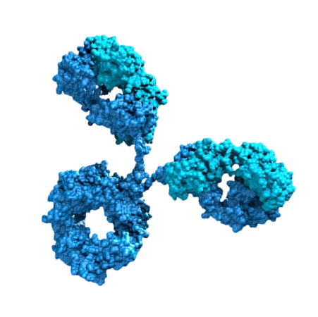
Phage display is a well-established and highly versatile technology for the discovery of binding molecules. By displaying antibody fragment libraries on filamentous phages, this approach enables efficient screening and enrichment of target-specific binders from highly diverse libraries.


At NBbiolab, our Phage Display VHH Discovery Service integrates high-quality library construction, optimized biopanning strategies, and advanced screening workflows to rapidly identify high-affinity nanobody candidates for research and therapeutic development.

When to Use Phage Display

Phage display is particularly suitable for:

Early-stage nanobody discovery

Screening against diverse antigen types (protein, peptide, cell surface targets)

Rapid enrichment of binders from large libraries

Projects requiring high library diversity

Initial binder identification prior to affinity maturation

Our Phage Display VHH Discovery Workflow
Deliverables

Clients will receive

High-affinity VHH sequences

Clone information and sequence data

Binding validation results

Expression-ready constructs

Detailed project report


Optional downstream services

Nanobody humanization

Affinity maturation

Functional characterization

Typical Project Timeline

Stage

Delivery

Time

Library construction

Phage library

~4 weeks

Screening

Binder sequences + report

~4 weeks

Total timeline: 6–8 weeks

The timeline above covers the yeast display library construction and screening stages of nanobody discovery. If camelid immunization is required, an additional 6–8 weeks may be needed.
Why Choose Our Phage Display VHH Discovery Service
High-Diversity Libraries
Library sizes up to 10⁹–10¹¹ ensure broad binder coverage
Optimized Biopanning Strategy
Customized panning conditions improve enrichment efficiency
Extensive Project Experience
Proven success across multiple target classes
Fast Turnaround Time
Efficient workflows reduce project timelines
FAQs
What are the phage display vector, host strain and helper phage adopted?

The phage display vector used is pComb3xss with HA and His tags; the host strain is TG1, and the helper phage is M13K07.

What are the quality control indicators for phage libraries?

Phage library quality control covers three aspects:
1. Library capacity calculated by transformants, reaching up to 10⁹ for phage libraries;
2. 100% insertion rate of target fragments;
3. Random selection of 48 clones for sequencing to evaluate library diversity.

Is nested PCR adopted for amplifying single-domain antibody genes for library construction? If yes, what are the amplification positions of primers in two rounds of nested PCR?

Nested PCR is used for single-domain antibody gene amplification in library construction. In the first round of nested PCR, the upstream primer targets the antibody signal peptide region and the downstream primer targets the CH2 region. In the second round, the upstream primer is located in the antibody FR1 region and the downstream primer in the hinge region.

What screening formats are available for phage display?

Common phage display screening formats include solid-phase screening, liquid-phase screening and cell-based screening.

Can phage display screen functionally active antibodies such as blocking antibodies?

Phage display is more inclined to screen binding antibodies, with inherent system limitations in screening functional antibodies (blocking, neutralizing and activating antibodies).

For screening blocking, neutralizing or activating antibodies, the positive control antibody can first be constructed into the phage vector to verify the applicability of the screening model and detection system. Formal screening can be launched only after system feasibility is confirmed.

Which sample form is adopted for screening and identification, phage supernatant or periplasmic protein?

Phage supernatant is preferred in the primary screening stage. Periplasmic protein is generally used for re-verification and activity identification of positive clones in the later stage.

Can monoclonal flow cytometry identification be performed in phage screening?

Flow cytometry-based binding identification of monoclonals with target cells is feasible. A phage-form positive control must be set in the experimental system to eliminate interference from background signals and non-specific binding.

See Our Wide Array of NANOBODY Services
Immunogen Design & Production
Camelid Immunization
Phage Display Nanobody Discovery
Functional Characterization
Yeast Display Nanobody Discovery
Premade Library Screening
Yeast Display Peptide Library Screening
Nanobody Discovery for ADC Development
Yeast Display Nanobody Discovery
Premade Library Screening
Yeast Display Peptide Library Screening
Nanobody Discovery for ADC Development
Related Services
Request a Quote